September 9, 2025
RNAscope & Xenium: 5 Powerful Tools Revolutionizing RNA Mapping
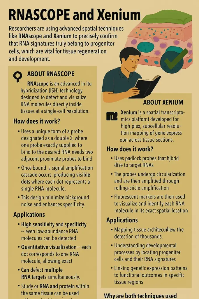
RNAscope and Xenium
Researchers are using advanced spatial techniques like RNAscope and Xenium to precisely confirm that RNA signatures truly belong to progenitor cells, which are vital for tissue regeneration and development.
About RNAscope
RNAscope is an advanced in situ hybridization (ISH) technology designed to detect and visualize RNA molecules directly inside tissues at a single-cell and even subcellular resolution.

How does it work?
- Uses a unique form of a probe designated as a double-Z, where one probe exactly supplied to bind to the desired RNA needs two adjacent proximate probes to bind.
- Once bound, a signal amplification cascade occurs, producing visible dots where each dot represents a single RNA molecule.
- This design minimizes background noise and enhances specificity.
Key Features:
- High sensitivity and specificity – even low-abundance RNA molecules can be detected.
- Quantitative visualization – each dot corresponds to one RNA molecule, allowing exact counting.
- Can detect multiple RNA targets simultaneously (multiplexing).
- Study of RNA and protein within the same tissue can be used with immunohistochemistry.
Applications:
- Validating gene expression data from bulk or single-cell RNA sequencing.
- Practical: The discovery of rare communities of cells (such as stem or progenitor cells).
- Understanding tissue organization and disease pathology, such as in cancer or neurological diseases.
About Xenium:
- Xenium is a spatial transcriptomics platform developed for high-plex, subcellular resolution mapping of gene expression across tissue sections.
How does it work?
- Uses padlock probes that hybridize to target RNAs.
- The probes undergo circularization and are then amplified through rolling-circle amplification.
- Fluorescent markers are then used to visualize and identify each RNA molecule in its exact spatial location.
- Multiple imaging cycles allow the detection of thousands of genes throughout the tissue in one experiment.
Key Features:
- Able to simultaneously identify thousands of distinct RNA targets due to its high multiplexing capability.
- Subcellular resolution – shows exactly where inside the cell each RNA molecule is located.
- Advanced software for data visualization and analysis, giving precise maps of gene expression.
- Can be combined with other techniques like Visium HD for broader tissue analysis followed by detailed zoom-in mapping.
Applications:
- Mapping tissue architecture in health and disease.
- Understanding developmental processes by locating progenitor cells and their RNA signatures.
- Studying tumor microenvironments and immune cell interactions.
- Linking genetic expression patterns to functional outcomes in specific tissue regions.
Why are both techniques used together?
- RNAscope provides a precise, targeted validation of a few specific RNA molecules, confirming their presence and location with high accuracy.
Xenium provides a broader, high-throughput view, mapping thousands of RNA species at once to show complex tissue patterns.
October 6, 2025
September 24, 2025
September 23, 2025
September 22, 2025
